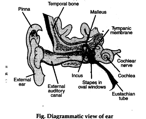
Each ear consist of three portions : (a) External ear, (b) Middle ear, © Internal ear.
(a) External ear : It comprises a pinna and external auditory meatus (canal).
(i) Pinna: It is a projecting elastic cartilage covered with skin. It outer ridge is called helix and the soft lower part is called lobule.
(ii) External Auditory Meatus : It is a tubular pas¬sage supported externally by cartilages and internally by bones. The canal is lined by ceru-minous glands.
(b) Middle ear: It includes the following:
(i) The tympanic membrane separates tympanic cavity from meatus, and is thin and semi-transparent.
(ii) The tympanic cavity is filled with air is connected with nasopharynx through eustachian tube, which equalizes the air pressure in the cavity.
(iii) There is a small flexible chain of three small bones called ear ossicles, malleus, incus and stapes.
(iv) The middle ear is connected with the inner ear through two small openings closed by the membranes. These openings are fenestra ovalis and fenestra rotunda.
© Internal ear: There is a bony cavity which contains perilymph, in which membranous labyrinth floats. The membranous labyrinth consists of three semicircular ducts, utricle, saccule, endolymphaticus and cochlea.
(i) Semicircular ducts : (1) There are present 3 semicircular ducts; the anterior, the posterior and the lateral semicircular ducts.
(2) They arise from the utricle.
(3) Each semicircular duct is enlarged at one end to give rise to a small rounded ampulla.
(4) Each ampulla contains a sensory patch of cells, the cristae. v
(5) Each crista consists of two kinds of cells, the sensory and supporting cells.
(ii) Utricle, Endolymphaticus and Saccule:
(1) The utricle is placed structure to which semicircular ducts are connected.
(2) The saccule is joined with the utricle by a narrow utriculosaccular duct. From this duct a long tube, the ductus endolymphaticus arises.
(3) Both utricle and saccule contain sensory patches, the maculae.
(4) Maculae comprises sensory and supporting cells.
(iii) Cochlea: (1) It is the main hearing organ which
is connected with saccule by a short ductus
reumiens.
(2) Internally it consists of three chambers, scala vestibuli, scala Temporal bone tympani and scala media.
(3) Both scala vertibuli and tympani are filled with perilymph and scala media is filled with endolymph.
(4) Scala media bears an upper membrane, Reissner’s membrane and lower basilar membrane, which has organ of corti.
(5) The sensory hairs project from hair cells
into scala media where arises cochlear nerve.